← Biology Resolution in Action

Case study · rheumatoid arthritis

Rheumatoid arthritis, read in patient-derived cells.

BioGPU compared fibroblast-like synoviocytes (FLS) from a rheumatoid arthritis patient with FLS from a healthy donor, using molecular-specific visualizers. The cellular system captured known RA biology, and the same experiments pointed to activities beyond it.

Category
Target Biology Resolution
Cellular system
Primary FLS · healthy donor vs RA patient
Readouts shown
18 visualizers in 8 panels
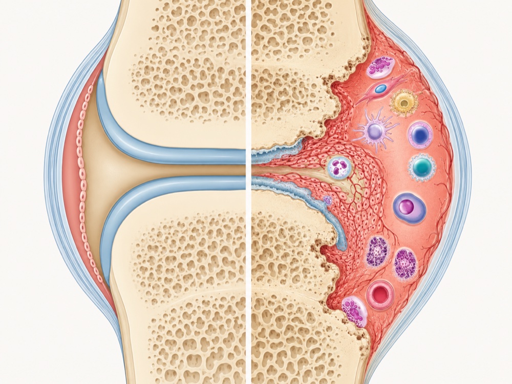
Illustration of a healthy joint (left) and a rheumatoid arthritis joint (right), with immune cells in the inflamed synovium
Healthy joint · RA joint (illustration). FLS line the synovium, beside chondrocytes (cartilage), osteocytes (bone) and immune cells.

Evidence · experimental images

Same visualizers, two donors, visible differences.

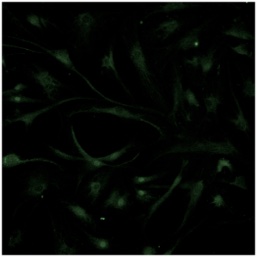
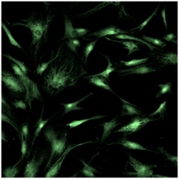
Choose an activity to compare the healthy donor (WT) with the RA patient (RA). Every pair is an experimental image from the study.

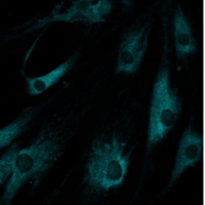
WT · healthy donorCSF2, healthy donor FLS
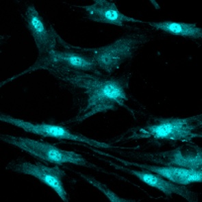
RA · patientCSF2, RA patient FLS
CSF2Inflammatory response

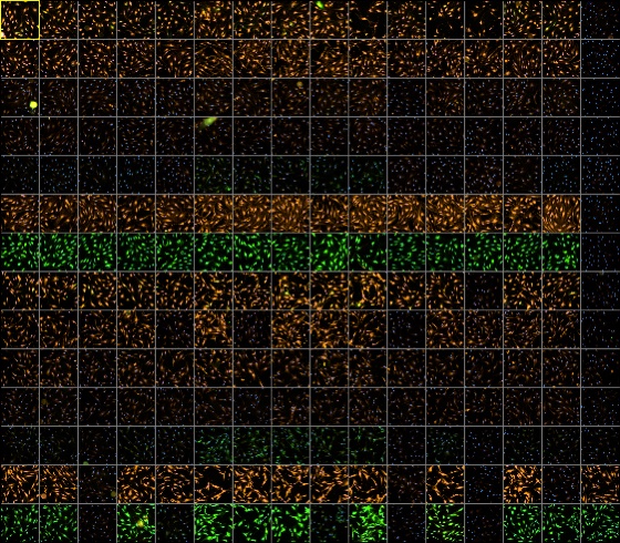
Activities were measured in parallel wells, not in one image.

BioGPU in action

From cellular events to statements AI can reason on.

  1. Plates of high-content imagesImageHigh-content imaging with molecular-specific visualizers
  2. Single-cell phenotypesSingle cellsEach cell's phenotype, then vectorized
  3. Clusters of single-cell phenotypesClusterCells grouped by shared visual patterns
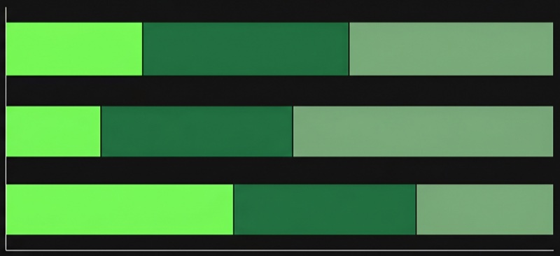
  4. Phenotype profiles as barsProfileHow each group shifts between conditions
  5. “Strong diffused cytoplasmic staining …”DescribePhenotype descriptions and scores, for biological interpretation

The Visual Biology Foundation Model recognizes and describes image-level phenotypes; biological reasoning across findings happens in AI agents.

One finding as a Visual Biology Token

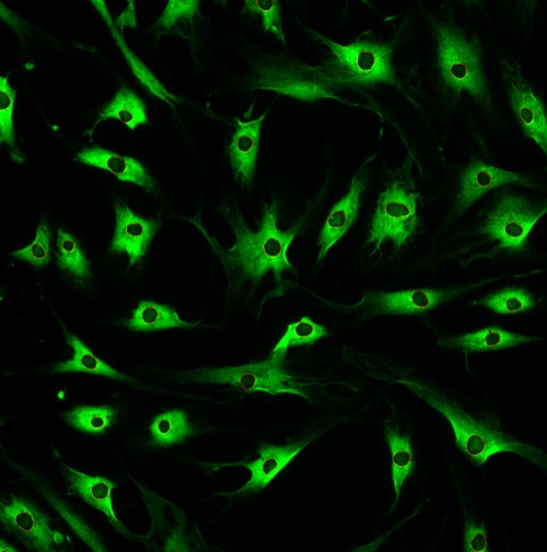
Src moves into the nucleus in RA cells.

Src, healthy donor FLS: cytoplasmic staining
WT · healthy donor
Src, RA patient FLS: nuclear staining
RA · patient

Image evidence · model description

Test cells show a dramatic shift in staining patterns compared to reference cells. The most striking change is a massive increase in cells displaying moderate diffuse nuclear staining with rounded dark nuclear holes accompanied by moderate granular cytoplasmic staining, indicating the emergence of a phenotype virtually absent in the reference group. Concurrently, there is a near-complete loss of cells exhibiting weak diffuse nuclear staining paired with moderate diffuse cytoplasmic staining, a phenotype that predominates in the reference population.

Biological meaning · interpretation

Non-phosphorylated Src translocated in RA-FLS cells from the cytoplasm to the nucleus. Nuclear, inactive, Src scaffolds the splicing machinery, converging with the alternative splicing hypothesis implied by SRPK1-SRSF activation.

Experimental contextSrc · primary FLS · healthy donor (reference) vs RA patient (test)

What this demonstrates

A patient's disease biology, captured and read in one cellular system.

Shown in this study
  • A patient-derived RA cellular system run on BioGPU beside a healthy donor.
  • Visualizers read inflammation, metabolism, citrullination, MAPK signaling, splicing, migration and mRNA translation in the same system.
  • Known RA biology appears in the images, and new activity patterns appear beside it.
  • The foundation model turns a phenotype shift into a description, kept with its images.
What it could inform · not shown here
  • Which targets to prioritize in disease-relevant cells.
  • A disease fingerprint that compounds can be read against.
  • Not shown: causality, therapeutic effect, or results across many donors.

See how a program runs → Explore cellular systems →

Resolve your target in disease-relevant cells.